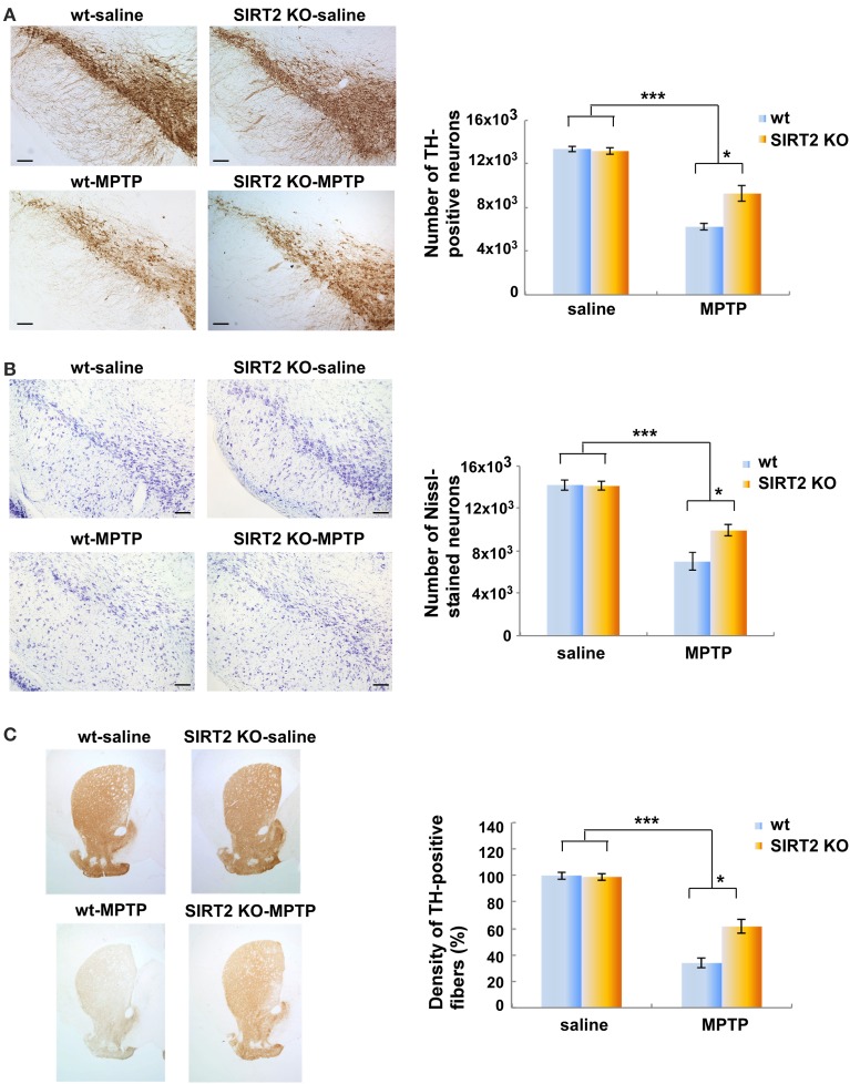
Figure 1.
Deletion of SIRT2 reduces the MPTP-induced nigrostriatal damage in mouse brains. (A) The left panel shows immunostaining of TH-positive neurons in the substantia nigra pars compacta (SNpc) in saline- or MPTP-dosed wt or SIRT2 KO mice. Scale bar represents 100 μM. The quantification on the right shows the number of SNpc neurons counted by the image analysis tool of NIS-Elements AR microscope software. Bars represent s.e.m (n = 4–5). Statistical analyses were carried out using Two-Way ANOVA. ***p < 0.001, MPTP vs. saline groups; *p < 0.05, SIRT2 KO-MPTP vs. wt-MPTP. (B) The left panel shows the Nissl staining of the neurons in SNpc of saline- or MPTP-dosed mice. Scale bar represents 100 μM. The quantification on the right shows the number of Nissl-stained neurons in SNpc counted by the image analysis tool of NIS-Elements AR microscope software. Bars represent s.e.m (n = 4–5). Statistical analyses were carried out using Two-Way ANOVA. ***p < 0.001, MPTP vs. saline groups; *p < 0.05, SIRT2 KO-MPTP vs. wt-MPTP. (C) The left panel shows the TH-positive striatal fibers in saline- or MPTP-dosed wt or SIRT2 KO mice. Quantification on the right shows the loss of striatal fibers in MPTP-dosed mice assessed by optical density (n = 4–5). Statistical analyses were carried out using Two-Way ANOVA. ***p < 0.001, MPTP vs. saline groups; *p < 0.05, SIRT2 KO-MPTP vs. wt-MPTP.

