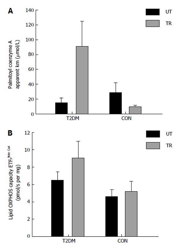
Figure 3.

Patients with type 2 diabetes (n = 5) and healthy control subjects (n = 3) performed eight sessions of one-legged high intensity training in two weeks. Each session consisted of ten one-minute exercise bouts at 60% of one-legged maximal oxygen uptake and > 80% of maximal heart rate, interspersed by one min rest. After completion of the training muscle biopsies (vastus lateralis) were obtained from the untrained (black bars) and the trained (grey bars) leg. The measurement mitochondrial OXPHOS capacity and substrate sensitivity was performed with malate, ADP and palmitoyl coenzyme A (titration: 5-100 μmol/L). A: Apparent Michaelis Menten constant Km for palmitoyl coenzyme A; B: Maximal OXPHOS capacity with the mentioned substrates. T2DM: Type 2 diabetes; CON: Control subjects; UT: Untrained; TR: Trained.
