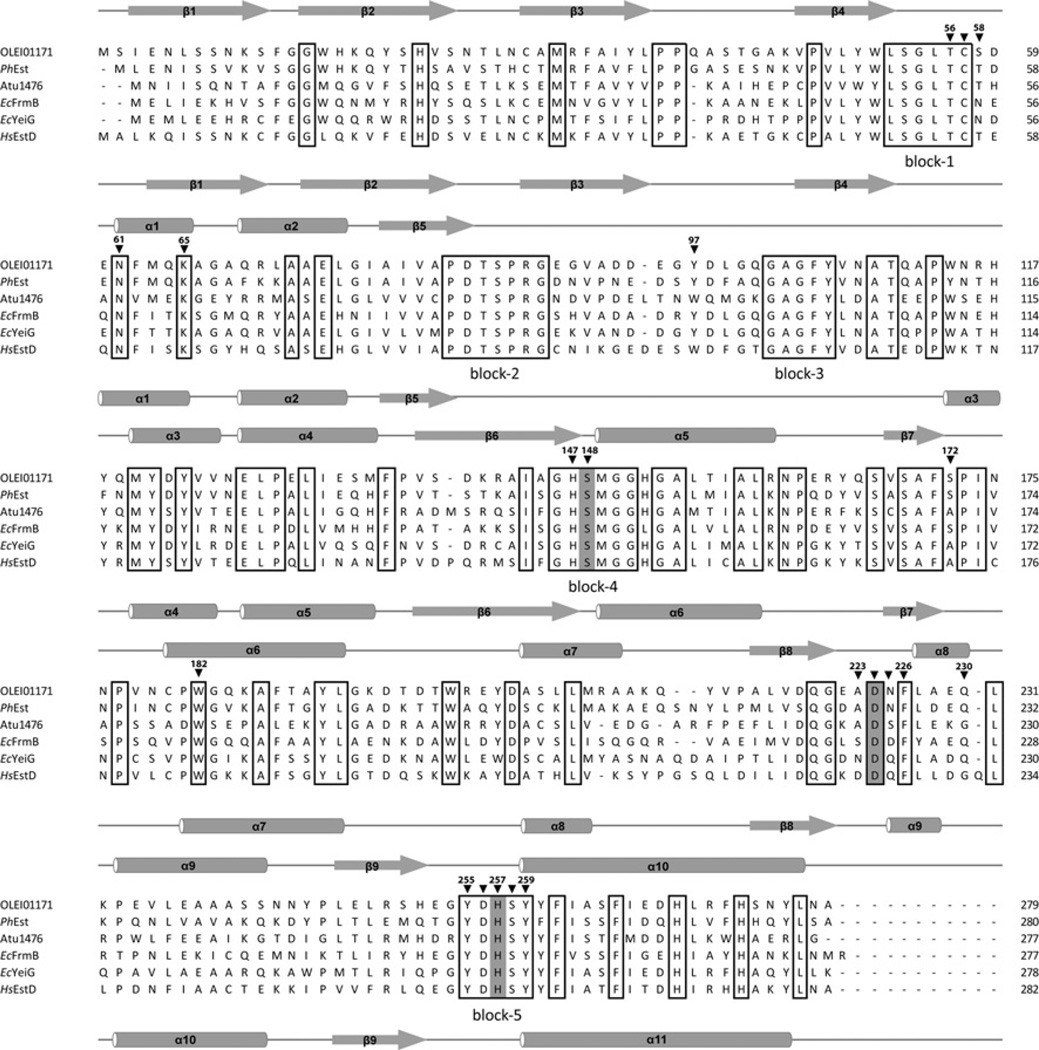
Figure 1. Structure-based sequence alignment of OLEI01171 and homologous esterases from bacteria and humans.
Residues conserved in all aligned proteins are boxed, and the residues constituting the serine hydrolase catalytic triad are highlighted in grey. The five large regions with conserved sequences are labelled ‘block-1’ to ‘block-5’ below the alignment. The OLEI01171 residues mutated in the present study are marked with black triangles above the alignment and numbered. The secondary structure elements derived from structures of OLEI01171 and human EstD are shown above and below the alignment respectively. The compared proteins are OLEI01171 (GenBank® accession number D0VWZ4), Ps. haloplanktis PhEst (GenBank® accession number Q3IL66), A. tumefaciens Atu1476 (GenBank® accession number A9CJ11), E. coli FrmB (EcFrmB; GenBank® accession number P51025) and YeiG (EcYeiG; GenBank® accession number P33018) and human EstD (HsEstD; GenBank® accession number P10768).

