
Figure 2.
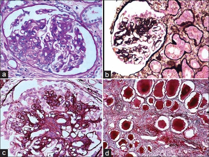
(a) Cellular variant of FSGS (PAS, ×200). (b) Segmental collapse of capillary tufts associated with podocyte hyperplasia and hypertrophy in a case of collapsing FSGS (Silver, ×200). (c) High-power view showing capillary collapse and podocyte hyperplasia and hypertrophy (Silver, ×400). (d) The tubulointerstitial compartment reveals moderate interstitial inflammation, tubular atrophy, and typical hyaline casts with scalloped edges in some of the tubular lumena (Silver, ×200)