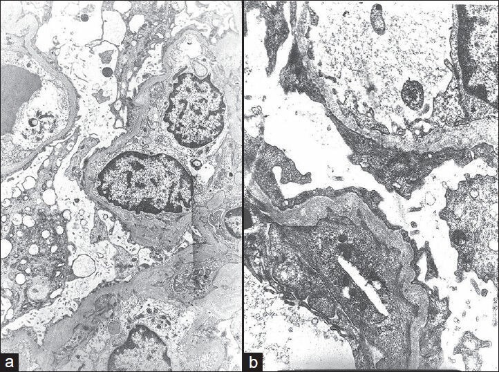
Figure 3.

(a) Electron micrograph showing extensive fusion of foot processes and marked cytoplasmic vacuolization of podocytes (electron microscopy, ×3,000). (b) Electron micrograph showing extensive fusion of foot processes, irregular thickening, and wrinkling of glomerular basement membrane (EM, ×10,000)
