
Figure 1.
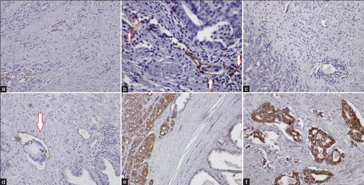
Detection of lymphatics by D2-40 immunostaining in prostate carcinoma (a-b). Note peritumoral lymphatic invasion (B, arrows). Limited LYVE-1 expression in prostate cancer (c-d). Note invasion of lymphatic vessel by tumor in D (arrow). Immunostaining for VEGF-C in prostate carcinoma (e–f). Strong cytoplasmic staining was present in the tumor cells, with extremely limited staining in the stroma and BPH cells (e). All microphotographs ×200 magnification