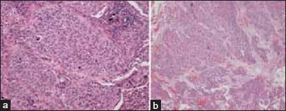
Figure 2.

(a) Photomicrograph of tumor resected from the left lower lobe showing a carcinoma with morphological features in keeping with metastatic transitional cell carcinoma. This interpretation was supported by immunohistochemical staining which showed characteristic expression of CK7, CK20, and nuclear staining for p63. Similar appearing metastatic carcinoma was also identified within hilar lymph nodes removed at the time of surgery (hematoxylin and eosin stain, ×100 original magnification). (b) Photomicrograph of tumor resected from the urinary bladder showing transitional cell carcinoma (hematoxylin and eosin stain, ×100 original magnification)
