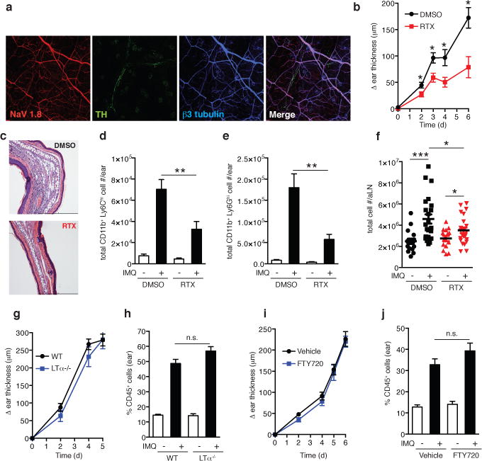
Figure 1. TRPV1+ nociceptor ablation attenuates skin inflammation and draining lymph node hypertrophy in the IMQ model.

a, Representative whole-mount confocal micrograph of normal ear skin from NaV1.8-TdT reporter mice (NaV1.8+ nociceptors, red) stained for β3-tubulin (peripheral nerves, blue) and tyrosine hydroxylase (TH, sympathetic nerves, green). b–f, The ear skin of vehicle treated controls (DMSO) or TRPV1+ nociceptor ablated (RTX) mice was treated with topical IMQ cream daily. (b) Ear thickness was measured relative to the contralateral ear at indicated time points (n=10–15 mice per time point; *, P < 0.02). (c) Representative histological sections of IMQ treated ears at day 6 stained by H&E. (d) Total inflammatory monocytes (n=10) and (e) total neutrophils in skin at day 3 (n=10; **, P < 0.005). (f) Total cell number in auricular lymph nodes at day 3 (n= 20; *, P = 0.01; ***, P < 0.001). g–j, IMQ was applied daily to (g,h) WT (n=10) and LTα −/− mice (n=6) or (i,j) vehicle treated (n=10) and FTY720 treated mice (n=10) and (g,i) ear swelling was measured at indicated time points. (h,j) The percentage of CD45+ leukocytes was determined in ear skin digests on day 6.
