
Extended Data Figure 9. Dermal DCs (DDCs) are found in close apposition to NaV1.8+ nociceptors in skin, NaV-DTA mice express reduced levels of key nociceptor markers, yet nociceptor deletion does not grossly affect the peripheral neural network in skin.
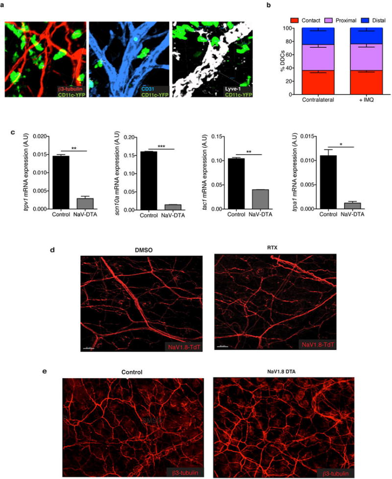
a, Representative confocal micrographs of CD11c-YFP mice stained for β3-tubulin, Lyve-1 (collecting lymphatics), and CD31 (blood and lymphatic endothelial cells). b, 3D quantification of DDC proximity to peripheral nerves in naïve and 6 hours post-IMQ treatment binned into contact (<0 um), proximal (0–7 um) and distal (>7 um) fractions as explained in the methods (n of DCs = 200). c, Total RNA from dorsal root ganglia (DRGs) (C1–C4) of littermate control and NaV1.8-DTA mice was isolated and levels of mRNA for trpv1 (TRPV1), scn10a (NaV1.8), tac1 (Substance P) and trpa1 (TRPA1) were determined relative to gapdh. This demonstrates the efficacy of the NaV1.8-DTA system and combined with the original reference characterizing the pain phenotype of these mice illustrates that a subset of peptidergic TRPV1+ nerve fibers is spared. d, Representative confocal micrograph of whole mount ear skin of Vehicle- and RTX-treated mice showing preserved nerve scaffold and e, representative confocal micrographs of whole mount ear skin of Control and NaV1.8-DTA mice showing preserved nerve scaffold. While DRGs showed a loss of the hallmark ion channels of these nerve subsets (Extended Data Fig. 1c and Extended Data Fig. 9c), surprisingly we still observed that RTX mice and NaV1.8-DTA mice maintain a meshwork of nerves in the skin.