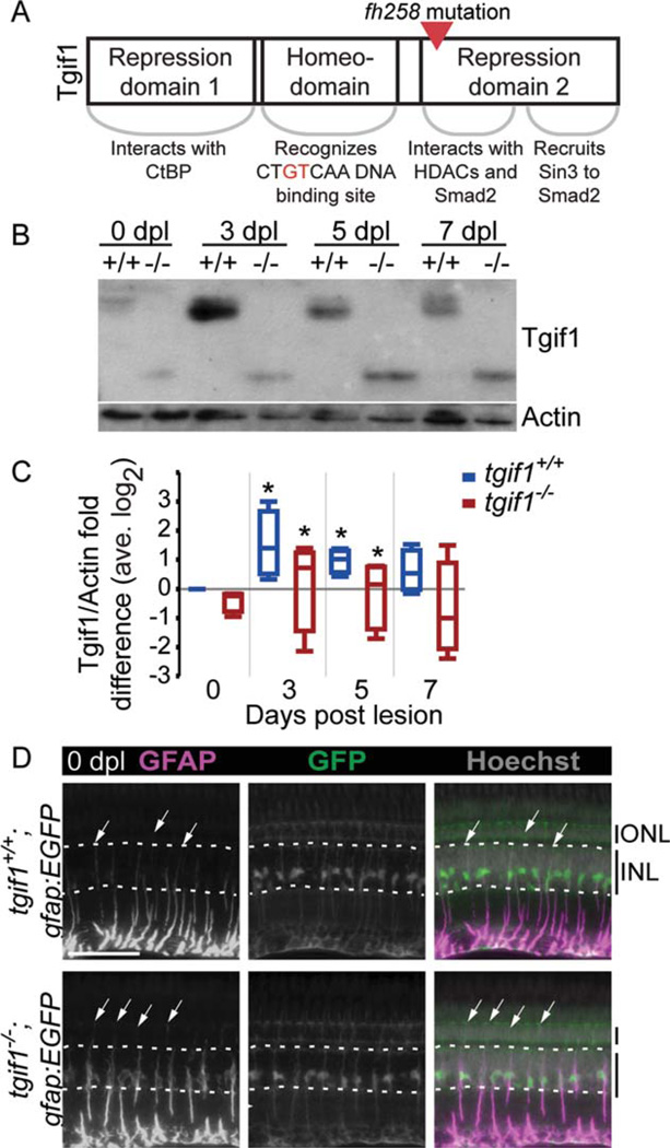
FIGURE 2.
Fish homozygous for the fh258 allele of tgif1 express a truncated Tgif1 and GFAP localization in Müller glia is altered. (A) Schematic of the Tgif1 protein and the point mutation in the fh258 allele. (B) Representative Tgif1 Western blot of whole retina extract from tgif1+/+ and tgif1−/− fish. (C) Densitometric analysis of Tgif1 protein in control and light lesioned retinas from tgif1+/+ and tgif1−/− fish normalized to actin and relative to 0 dpl tgif1+/+ (retinas pooled from two fish, n = 4 replicates). * P<0.05 relative to 0 dpl of the same genotype, Student t-test. (D) GFAP immunolocalization (first column) in the unlesioned retina of tgif1+/+;gfap:EGFP (top) and tgif1−/−;gfap:EGFP (bottom) fish expressing GFP only in the Müller glia. We used the same exposure time and did not alter the GFAP images postcapture. Arrows = top of GFAP distribution in individual Müller glia; dotted white lines delineate the base and top of the INL. Abbreviations as in Fig. 1. Scale bar = 50µm.

