
Figure 2. A basement membrane peroxidase forms the collagen IV sulfilimine bond.
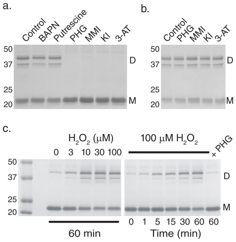
(a) Coomassie blue stained gel after SDS-PAGE of NC1 hexamers isolated from PFHR-9 cells grown in the presence of β-aminopropionitrile (BAPN; 500 μM), putrescine (2.5 mM), phloroglucinol (PHG; 50 μM), methimazole (MMI; 1 mM), 3-aminotriazole (3-AT; 10 mM), and potassium iodide (KI; 10 mM). Collagen IV NC1 hexamer from untreated cells (control) is shown for comparison. Gel is representative of 5 independent experiments. (b) PFHR-9 basement membrane was allowed to form normally, isolated, and treated with phloroglucinol (PHG; 50 μM), methimazole (MMI; 1 mM), potassium iodide (KI; 10 mM), and 3-aminotriazole (3-AT; 10 mM) for 24 hours at 37°C. Collagen IV NC1 hexamer was isolated and underwent SDS-PAGE and Coomassie blue staining to visualize sulfilimine cross-link content. (c) Coomassie blue stained gel after SDS-PAGE of NC1 hexamers after reacting uncross-linked PFHR-9 basement membrane with H2O2 at varying concentrations for 1 hour (left panel) or for varying durations with 100 μM H2O2 (right panel) in 1X PBS (phosphate buffered saline; 150 mM NaCl, 10 mM sodium phosphate, pH 7.4). The gel is representative of 8 independent experiments. D represents NC1 cross-linked dimeric subunits, while M denotes uncross-linked monomeric subunits. Full gel images are provided in Supplementary Fig. 13.