
Figure 3. Analysis of sg mRNAs produced by MA104 cells infected with SHFVic virus.
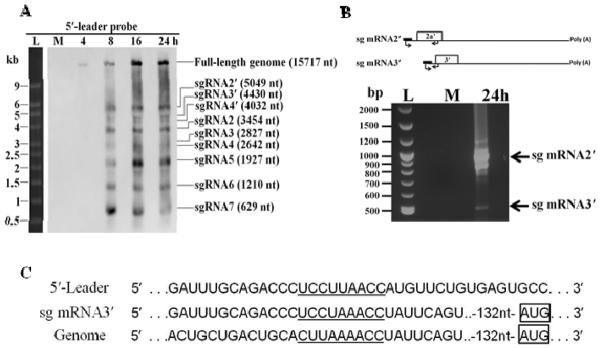
(A) Northern blot analysis of viral sg mRNAs. MA104 cells were mock infected (M) or infected with SHFVic virus at an MOI of 1 and total RNA was extracted at 4, 8 16 or 24 h after infection. Cell RNA (1 μg) was electrophoresed on a 1% denaturing agarose gel, transferred to an Hybond-N+ membrane and hybridized with a DIG-labeled 5′ leader RNA probe. The sg mRNAs were identified based on size. (B) Top-Diagram indicating the positions of the RT-PCR primers used. Bottom- Cell RNA was extracted from MA104 cells 24 h after infection with SHFVic virus at an MOI of 1 and subjected to RT-PCR. The PCR products were separated on a 2% agarose gel. The ~1000 and ~500 bp DNA bands were excised and the extracted DNA was TA cloned and sequenced. (C) The junction sequence for sg mRNA 3′ obtained from the ~500 bp band was aligned with the genome and 5′ leader sequences.