
Figure 6.
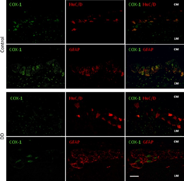
Double immunofluorescence of COX-1 (green)-HuC/D (red neurons) or -GFAP (red glial cells) in myenteric ganglia of colonic specimens from control subjects and DD patients. In control tissue, COX-1 is expressed in most of HuC/D+ neurons, in the nuclei of some myocytes of longitudinal layer (LM), and scarcely in GFAP+ glial cells. In samples of DD colons, COX-1 expression is reduced in HuC/D+ neurons, as demonstrated by merged COX-1-HuC/D images. Bar: 50 μm.