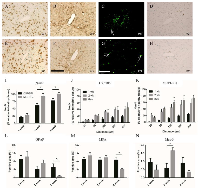
Figure 2.
Neuroinflammation was reduced in MCP-1 KO mice. Paraffin embedded brain sections were stained for NeuN to reveal near-implant neurons. MCP-1 KO (E) tissue showed an increased density of neurons when compared to WT (A) tissue. Quantification of NeuN positive area (I) showed that there was a statistically significant increase in neuronal density at 2 and 8 wks in the KO mice. Elliptical regions were used to quantify the amount of positive stain at distances from the implant interface in WT (J) and KO mice (K). Neuronal density was increased in the KO mice at all distances from the implant cavity (* in K compare WT and KO at the same time and distance). Reactive astrocytes were visible around the wound cavity 1, 2, and 8 wks in both WT (B) and KO (F) mice. The extent of reactive gliosis was reduced in KO mice at 8 wks (L), when quantified using MetaMorph. The presence of MSA was used to measure the extent of BBB leakage. Bright, punctuate areas of fluorescence, visible in all sections but highlighted by white arrows in C, and G, indicate unperfused vessels and were excluded from analysis. Both WT (C) and KO (G) tissue showed extensive leakage at 1 wk which gradually resolved over time (M). At 8 wks, the amount of BBB leakage in KO tissue was less than in WT tissue, which indicates greater BBB integrity and an improved FBR. Tissue sections were stained with Mac-3 to show local macrophage/microglia accumulation. WT mice showed consistent macrophage/microglia presence in WT mice from 1 to 8 wks (D, H, N). Macrophage accumulation peaked at 2 wks after implantation in the KO mice. Scale is 100 μm in F and G, * indicates p < 0.05 using Student’s t test, n is 6 for each point and data is presented ± SEM.

