Fig. 3.
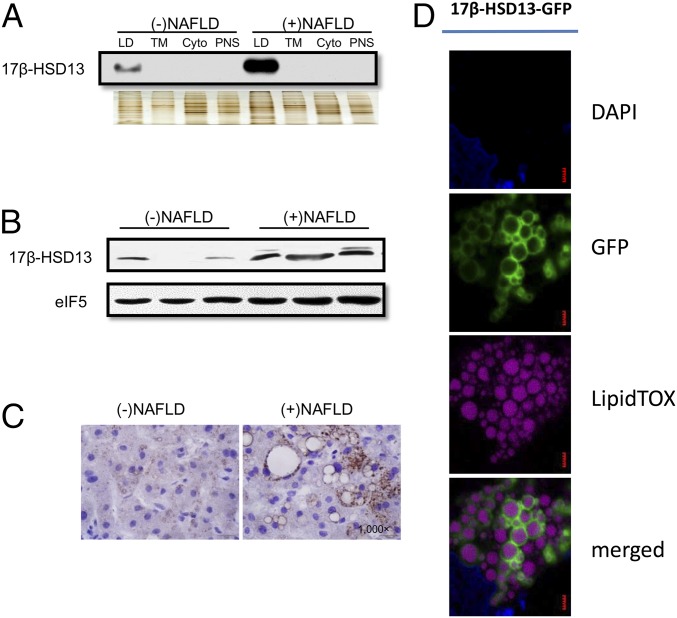
LD surface localization of 17β-HSD13 in human liver and cultured hepatocytes. (A) Western blot analysis showing that 17β-HSD13 was markedly up-regulated in LD fraction of NAFLD. Silver staining served as an even loading control. (B) Immunoblot assay using whole liver lysates demonstrating that 17β-HSD13 was significantly up-regulated in fatty livers. (C) Immunostaining of 17β-HSD13 showing that 17β-HSD13 was localized at the surface of LDs in human livers. Note: More intense staining of 17β-HSD13 in fatty liver than control liver. (D) LD surface localization of GFP-tagged 17β-HSD13 (17β-HSD13–GFP) in Huh7 cells. Green: 17β-HSD13-GFP; Mauve: LipidTOX Deep Red; Blue: DAPI. (Scale bar, 1 µm.)

