Fig. 3.
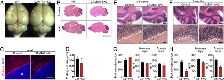
Degeneration of Purkinje cells and cerebellar atrophy in CAMTA1-nKO mice. (A) Gross morphology of brains from adult WT and CAMTA1-nKO mice. (B) H&E stain of sagittal sections of brain from 3-wk-old (Upper) and adult (Lower) WT and CAMTA1-nKO mice. (Scale bar, 400 μm.) (C) Calbindin-D28K immunohistochemical staining (red) of Purkinje cells in the cerebellum of 6-mo-old CAMTA1-nKO and littermate control mice. Purkinje cells are uniformly organized in the cerebellum of the control mouse but are reduced and disarrayed in the cerebellum of the CAMTA1-nKO mouse. Nuclei are stained with DAPI (blue). gl, granular layer. (Scale bar, 100 μm.) (D) Quantification of the size of calbindin-positive Purkinje cells in the cerebellum (n = 3). (E and F) H&E staining of sagittal sections of the brain from CAMTA1-nKO and littermate control mice at age 2–3 wk (E) and 3 mo (F). White arrows point to Purkinje cells. (Scale bar, 400 μm Upper, 40 μm Lower.) n = 6; *P < 0.05. gl, granular layer; ml, molecular layer. (G and H) Quantification of the Purkinje cells of CAMTA1-nKO and littermate control mice at age 2–3 wk (G) and 3 mo (H). Purkinje cell number is not reduced in CAMTA1-nKO mice at age 2–3 wk but is decreased at age 3 mo along with reductions in the width of the molecular layer and granular layer. Data are presented as mean ± SD.

