Fig. 5.
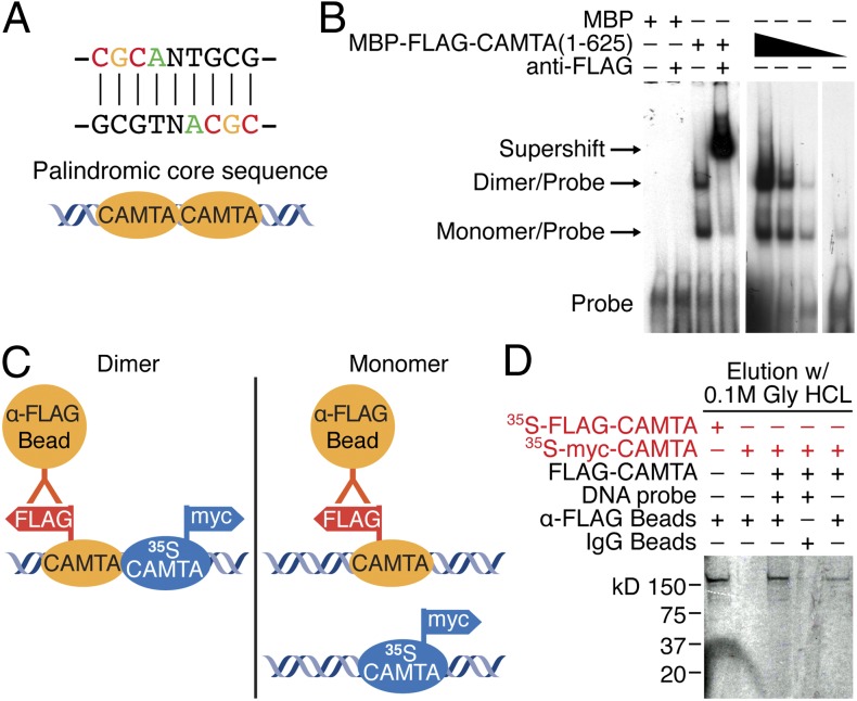
Dimerization of CAMTA on the palindromic core sequence. (A) Schematic of CAMTA dimers on the palindromic core sequence. (B) Gel mobility shift assay consisting of recombinant protein of the N terminus (amino acids 1–625) of CAMTA (MBP-FLAG-CAMTA) incubated with its DNA-binding site generates two bands on the gel. Both bands were supershifted by anti-FLAG antibody. When decreasing amounts of CAMTA protein are added (designated by the black triangle), the ratio of the intensity of the upper band to the intensity of the lower band is decreased. The higher band is a complex of a CAMTA homodimer and DNA probe. The lower band is a complex of a CAMTA monomer and DNA probe. (C) Schematic of the in vitro pull-down assay of dimerization of CAMTA. (D) Pull-down assay showing 35S-labeled, myc-tagged CAMTA incubated with FLAG-CAMTA and the DNA core sequence could be pulled down with anti-FLAG beads, whereas 35S-labeled, myc-tagged CAMTA alone or with DNA was not pulled down by anti-FLAG beads.

