Fig. 4.
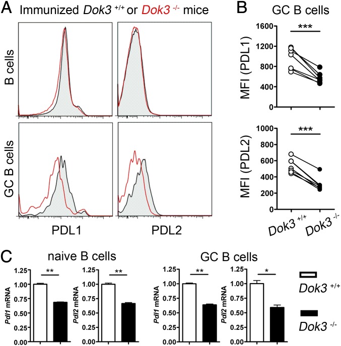
DOK3 deficiency leads to impaired PDL1 and PDL2 expression in B cells. (A) Representative histogram of protein expression levels of PDL1 and PDL2 on splenic B and GC B cells in WT and Dok3−/− mice at day 10 after immunization. (B) Cumulative flow cytometry analyses of mean fluorescence intensity (MFI) for PDL1 and PDL2 protein expression on GC B cells as shown in A. (C) Quantitative real-time PCR analyses of PDL1 and PDL2 mRNA levels in naïve B cells from unimmunized or GC B cells from immunized WT and Dok3−/− mice. mRNA level is normalized to that of GAPDH. *P < 0.05; **P < 0.01; ***P < 0.001.

