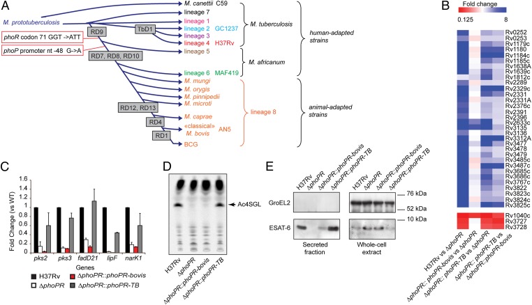
Fig. 1.
The phoPR allele from animal-adapted and M. africanum L6 strains is deficient. (A) Schematic global phylogenetic tree of the MTBC (1, 2). The length of the branches does not correlate with phylogenetic distance. Names of the strains used in this study are indicated. (B) Genome-wide transcriptional profiles of WT M. tuberculosis H37Rv, ΔphoPR mutant, and phoPR-bovis– or phoPR-TB–complemented strains. Fold-change values from individual probes for each gene were averaged. Those genes showing a statistically significant average fold-change > 2 or < 0.5 in the WT or the phoPR-TB–complemented strains relative to the ΔphoPR mutant were selected as positively or negatively regulated by PhoP, respectively. (C) qRT-PCR analysis of expression of main reporter genes of the PhoP regulon. (D) TLC analysis of lipids extracted from [14C] propionic acid-labeled cultures. The position of the major SL (the tetra-acylated sulfoglycolipids, Ac4SGL) is highlighted (arrow). (E) Immunoblot of secreted and whole-cell fractions probed with ESAT-6– or GroEL2- (used as a lysis control) specific antibodies.

