Fig. 5.
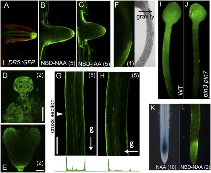
Distribution of fluorescent auxin mimics the native auxin gradient. (A–C) DR5::GFP expression and fluorescent signals of NBD-auxins in lateral root primordia. (D and E) NBD-NAA distribution in preglobular embryos and heart-stage embryos. (Scale bars in A–E, 20 μm.) (F–H) Gravity-stimulated 3-d-old etiolated hypocotyls were treated with 1 μM NBD-NAA for 15 min (F) or 5 μM NBD-NAA (G and H) for 30 min while maintaining the unidirectional stimulus of gravity (g). Arrowhead indicates the cross section location. (Scale bar in G and H, 200 μm.) (I and J) Distribution of NBD-NAA in 4-d-old etiolated hypocotyls of wild type and pin3 pin7 mutant. Agarose gel (0.1%, 0.2 µL) containing NBD-NAA (80 µM) was applied on the shoot apex and then incubated for 4 h in the dark. (K and L) Rice DR5::GUS roots were incubated with 10 μM NAA for 24 h (K). Distribution of NBD-NAA in rice roots (L). The values in parentheses indicate the concentrations of chemicals (μM).

