
Figure 6. Hb9 gain of function in ap neurons induces robo2 expression and a robo2-dependent lateral shift.
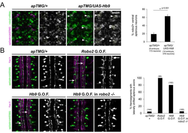
A, Left: Fluorescent in situ for robo2 mRNA (green) in Stage 15 embryos. Anterior is up. The ventral ap neurons are labeled in magenta and circled in the single channel images. Wild-type embryos express little robo2 in the ap neurons, whereas many ventral ap neurons express robo2 when Hb9 is present (arrowheads). A, Right: The percentage of ventral ap neurons expressing robo2 is shown. Hb9 gain of function results in a significant increase compared to controls (p<0.001, Student’s t-test). Error bars = s.e.m. B, Left: Stage 17 embryos stained for FasII (magenta) and GFP (green), which labels the ap axons. Over-expression of robo2 or hb9 in ap neurons shifts their axons laterally (arrows). Hb9 over-expression in robo2 mutants does not induce a lateral shift phenotype. B, Right: The percentage of hemisegments in which ap axons project along the intermediate or lateral FasII tracts is shown. Numbers of hemisegments scored are indicated in parentheses. Scale bars represent 10 μm. apTMG/+ denotes apGal4,UAS-TauMycGFP/CyO. Robo2 G.O.F. denotes UAS-HARobo2.T1/apGal4, UAS-TauMycGFP. Hb9 G.O.F denotes UAS-Hb9/apGal4,UAS-TauMycGFP. Hb9 G.O.F. in robo2 −/− denotes robo2123,UAS-Hb9/robo233, apGal4; UAS-TauMycGFP/+.