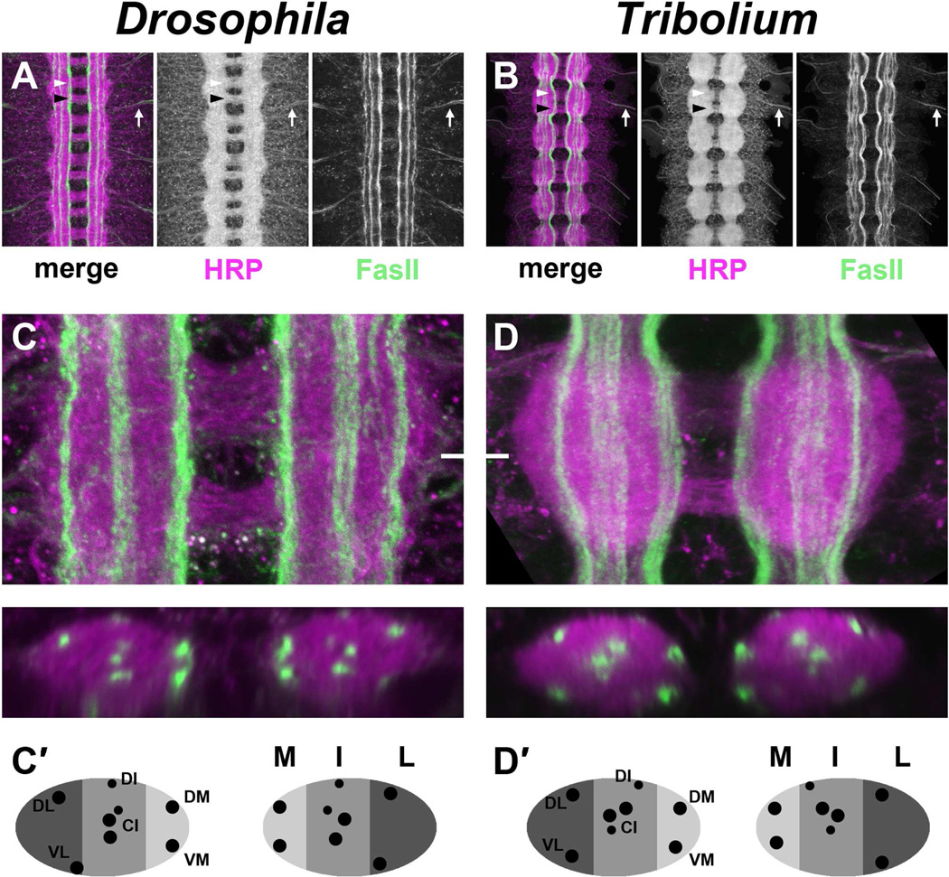
Figure 4. Conservation of CNS architecture in Drosophila and Tribolium.
A–B. Dissected ventral nerve cords from a stage 16–17 Drosophila embryo (A) and 4–5 day old Tribolium embryo (B) stained with antibodies against HRP (magenta; detects all axons in the CNS) and the longitudinal pathway marker FasciclinII (FasII; green). Both insects exhibit a ladder-like arrangement of axon pathways in the CNS consisting of bilateral longitudinal connectives and two commissures per segment. Anterior and posterior commissures in a single segment are indicated by white and black arrowheads, respectively. Individual FasII-positive axon pathways are visible in both species, and they do not cross the midline. Peripheral nerves are labeled by both anti-HRP and anti-FasII in each species as they exit the CNS (arrows).
C–D. Single abdominal segments of Drosophila (C) and Tribolium (D) embryos stained as in A–B. Maximum confocal projections through the entire neuropile are shown above; optical cross sections taken at the level of the white hash mark are shown below.
(C′,D′). Schematic representation of cross sections shown in C,D denoting medial (M, light gray), intermediate (I, medium gray) and lateral (L, dark gray) regions of the neuropile and showing relative positions of individual longitudinal pathways (black dots).
C–C′. FasII-positive pathways form in three distinct zones within the Drosophila neuropile. At least eight distinct FasII pathways can be seen in cross section: the dorsal medial (DM) and ventral medial (VM) pathways form within the medial (M) zone; three central intermediate (CI) and one dorsal intermediate (DI) pathway form within the intermediate (I) zone; the dorsal lateral (DL) and ventral lateral (VL) pathways form within the lateral (L) zone. Pathway nomenclature is after Landgraf et al 2003.
D–D′. Similar arrangement of axon pathways in the Tribolium embryonic CNS. Distinct FasII-positive pathways are visible at medial, intermediate, and lateral positions. The same number of individual pathways can be seen in cross section at equivalent dorsoventral and mediolateral locations to those in Drosophila. In this and all subsequent figures, Tribolium embryos are shown scaled to 75% original size for easier comparison with the smaller Drosophila embryos.

