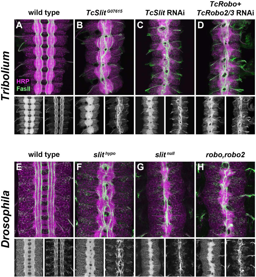
Figure 5. Slit/Robo signaling mediates midline repulsion in Tribolium.
A–D. 4–5 day old Tribolium embryos stained with anti-HRP (magenta) and anti-TcFasII (green). Small panels below show individual HRP (left) and FasII (right) channels.
A. Wild type Tribolium embryo. The ladder-like axon scaffold is formed by segmentally repeated pairs of commissures and bilateral longitudinal connectives. Distinct FasII-positive pathways are visible, and they do not cross the midline.
B. In TcSlitG07615 embryos, commissures are fused and FasII-positive axons linger at the midline, similar to the phenotype seen in Drosophila hypomorphic slit mutants (F). Some longitudinal pathways still form relatively normally in these embryos.
C. Embryos from mothers injected with dsRNA targeting TcSlit display a strong midline collapse phenotype. The width of the axon scaffold is reduced and longitudinal pathways do not form properly, as many FasII-positive axons enter the midline and fail to leave.
D. Embryos in which TcRobo and TcRobo2/3 are targeted in combination phenocopy the TcSlit knockdown phenotype.
E–H. Stage 16–17 Drosophila embryos stained with anti-HRP (magenta) and mAb 1D4 (anti-FasII; green).
E. In wild type Drosophila embryos, the longitudinal connectives are well separated and connected by two commissures per segment. Three distinct FasII-positive pathways can be seen on either side of the midline; they do not cross the midline.
F. In hypomorphic slit mutants, the connectives are closer together, commissures thicken and begin to fuse, and FasII-positive axons cross the midline in every segment.
G. In slit null mutants, all axons collapse at the midline, reflecting a complete absence of midline repulsion.
H. Double mutant robo,robo2 embryos display a slit-like midline collapse phenotype.

