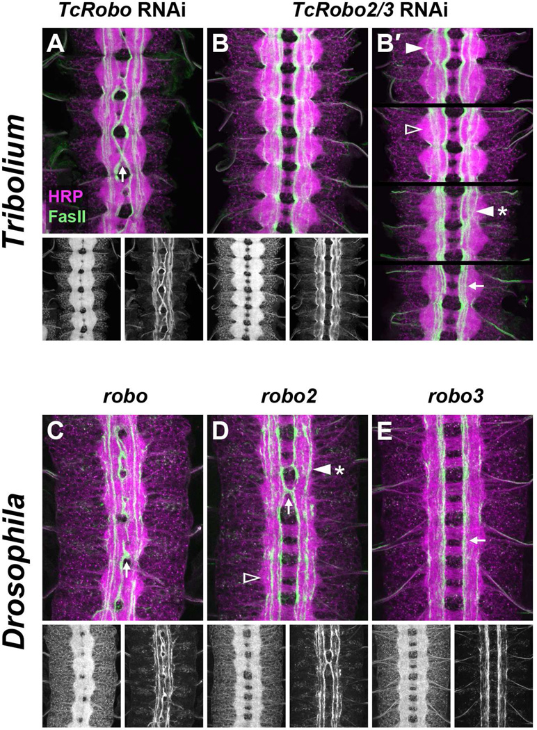
Figure 6. Tribolium Robo receptors exhibit specialized axon guidance functions.
Tribolium (A,B) and Drosophila (C–E) embryos prepared as in Figure 5.
A. TcRobo knockdown embryo. Commissures are thickened and FasII axons from medial pathways ectopically cross the midline (arrow). Intermediate and lateral longitudinal pathways are formed relatively normally.
B. TcRobo2/3 knockdown embryo. The axon scaffold has an overall normal appearance, and no FasII-positive axons cross the midline. Longitudinal pathways are disorganized and often fail to form at their normally stereotyped positions.
B′. Partial confocal projections from four different TcRobo2/3 knockdown embryos displaying longitudinal pathway defects: missing lateral pathways (empty arrowhead); FasII-positive axons wandering between longitudinal pathways (arrowhead with asterisk); intermediate and lateral pathways compressed into medial zone (arrow). In a minority of segments, intermediate and lateral pathways form relatively normally (solid arrowhead). Projections were made from 8–15 0.2 µm confocal sections and include all dorsal pathways in their entirety while excluding the majority of ventral pathways.
C. robo mutant embryo. As in TcRobo knockdown embryos (A), thickened commissures and ectopic midline crossing of FasII axons can be seen (arrow).
D. robo2 mutant embryo. Although the axon scaffold appears relatively normal, medial FasII axons can occasionally be seen crossing the midline (arrow). In addition, lateral FasII pathways are often absent (empty arrowhead) or fuse with intermediate pathways (arrowhead with asterisk).
E. robo3 mutant embryo. The axon scaffold appears normal, and no ectopic crossing is detectable. Intermediate FasII pathways shift towards the midline and fuse with medial pathways (arrow).

