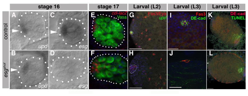
Figure 2. Loss of hub marker expression during larval development in esgshof males.

DIC images of RNA in situ for upd (A,B) or esg (C,D) in control (A, C) and esgshof (B, D) stage 16 embryonic gonads (outlined, arrowheads). Gonads from cdi-lacZ (E) and cdi-lacZ; esgshof (F) stage 17 embryos (outlined) stained for Vasa (green), β-galactosidase (red), DAPI (blue). Larval L2 updGAL4, UAS-GFP (G) and updGAL4, UAS-GFP; esgshof (H) gonads stained for Fas3 (red, outline), Eyes Absent (Eya, red), GFP (green), DAPI (blue). Note loss of GFP expression in (H), despite residual Fas3. Larval L3 control (I) and esgshof (J) gonads stained for Fas3 (red), E-cad (green), DAPI (blue). Larval L3 updGal4, UAS-GFP (K) and updGal4, UAS-GFP; esgshof (L) gonads stained for E-cadherin (red, asterisk), DAPI (blue), and TUNEL assay for apoptotic cells (green).
