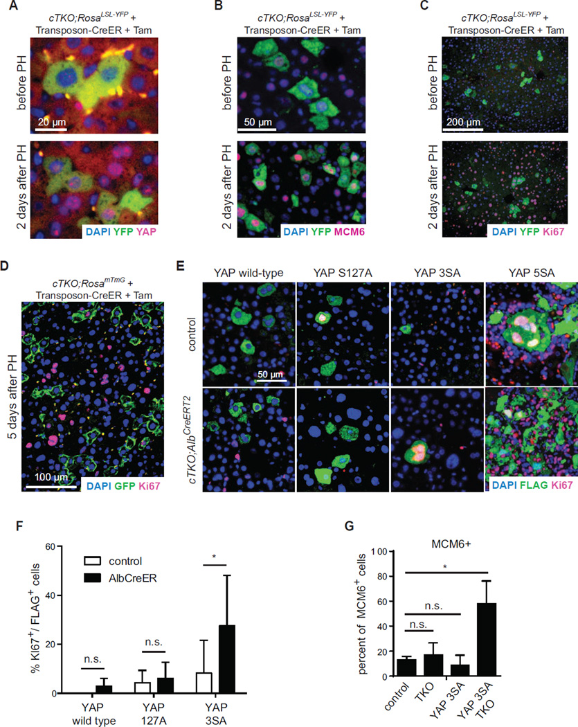
Figure 5. YAP activation is sufficient to revert the cell cycle arrest in TKO livers.
(A–C) Immunofluorescence analysis of YFP (green) together with endogenous YAP (A), MCM6 (B), or Ki67 (C), respectively, after 2/3rd partial hepatectomy (PH) 2 weeks after Tam in CreER transposon-injected cTKO;RosaLSL-YFP mice. (D) Immunofluorescence staining of GFP (green) and Ki67 (red) 5 days after 2/3rd PH, 2 weeks after Tam in CreER transposon-injected cTKO;RosamTmG mice. (E) Immunofluorescence analysis of YAP (Flag, green) and Ki67 (red) following hydrodynamic tail vein injection of a transposon expressing either wild type YAP or constitutively active variants of YAP (YAPS127A, YAP3SA, or YAP5SA) into control or arrested TKO livers (transposon injection 1 week after 50 µg Tam, livers analyzed 2 weeks after Tam). (F) Quantification of Ki67 positive YAP-transfected hepatocytes. Data represented as mean +/− SD. (G) Quantification of MCM6 positive hepatocytes following YAP3SA expression in controls or arrested TKO livers (transposon injection 1 week after 50 µg Tam, livers analyzed 2 weeks after Tam). Controls, Flag negative cells in control livers; TKO, Flag negative cells in arrested TKO livers; YAP3SA, Flag positive cells in control livers; YAP3SA TKO, Flag positive cells in arrested TKO livers.

