Figure 2.

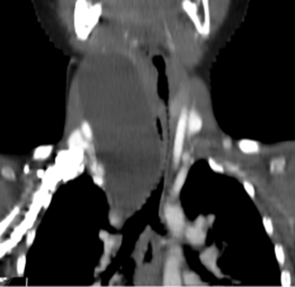
Patient B.K., 3-year-old boy with growing cyst on the neck. Contrast-enhanced CT, MPR in coronal plane: tubular esophageal duplication in the neck and mediastinum, clinically asymptomatic compression of the trachea.

Patient B.K., 3-year-old boy with growing cyst on the neck. Contrast-enhanced CT, MPR in coronal plane: tubular esophageal duplication in the neck and mediastinum, clinically asymptomatic compression of the trachea.