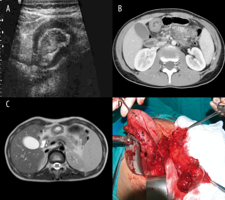
Figure 5.
Patient E.S., clinical signs of acute pancreatitis. (A) Sonographic image of thick-walled cyst in the prepyloric region of the stomach, lined with polypomatous hypertrophic mucosa. (B) Contrast-enhanced CT: thick-walled duplication cyst of the prepyloric region of the stomach with heterotopic foci of pancreatic tissue; accessory pancreas between the pancreas and stomach. (C) MRI image of the duplication cyst. (D) Intraoperative image.

