
Figure 4. Neutrophils modulate the capacity of the hematopoietic niche to attract and retain immature leukocytes.
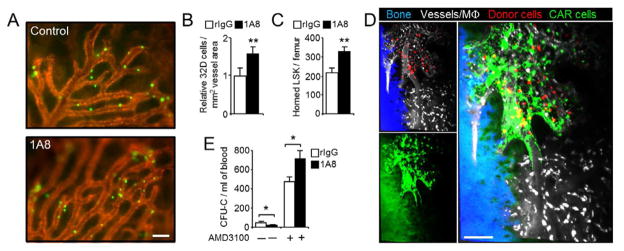
(A) Representative micrographs showing homed 32D cells (green) and BM microvessels (red) in control and 1A8-treated mice. Scale bar, 100 μm.
(B) Quantification of homed cells per vessel area in the BM of control (rIgG) or neutrophil-depleted (1A8) mice. Values were obtained from the experiments illustrated in (A). n =4–5 mice.
(C) Relative number of donor-derived DsRed+ LSK cells that home into the BM of control (rIgG) or neutrophil-depleted (1A8) mice, as determined by flow cytometry. n=5–6 mice.
(D) DsRed+ BM-derived donor cells (red) home exclusively into areas of the BM enriched in CXCL12-producing cells (green; background green on the left corresponds to autofluorescent bone). Vessels and macrophages (white) are visualized with fluorescent dextran, and bone (blue) was imaged by second harmonic generation (See also Movie S1). Small panels on the left are shown merged on the right panel. The micrograph is representative of four Cxcl12-Gfp mice imaged by multiphoton microscopy. Scale bar, 100 μm.
(E) Mobilization of control (rIgG) or neutrophil-depleted mice (1A8) with AMD3100. n=5–6 mice.
Data are shown as mean ± s.e.m. *P<0.05, **P<0.01.