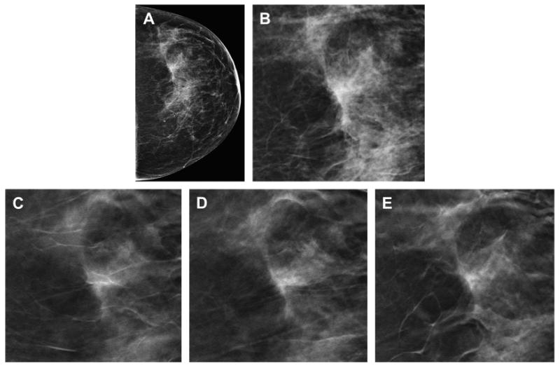
Fig. 1.

Reduction in false-positive callbacks with DBT. The DM CC view (A) demonstrates focal asymmetry with a suggestion of architectural distortion in the slightly lateral breast. A cropped, enlarged view of the DM focal asymmetry (B) better demonstrates the area of possible distortion. Multiple in-plane 1-mm reconstructed slices (C–E) from the DBT clearly show that the focal asymmetry seen on the two-dimensional DM study is caused by tissue superimposition rather than a clinically significant finding.
