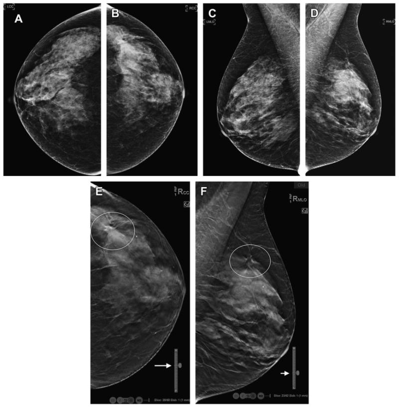
Fig. 10.

DBT localization tools. The routine, DM screening mammogram (A–D) shows no definite abnormality. On the DBT CC view (E) there is an area of architectural distortion in the lateral breast that is localized in the superior portion of the reconstructed stack of slices (Slice: 39/60 and close to the “H” or head, cranial aspect of breast as shown on vertical localizer marker, arrow). Now knowing were to search in the MLO DBT reconstructed slices (F), a very subtle area of distortion is seen in the superior and lateral aspect of the MLO stack (Slice: 23/62; closer to “L” or lateral side of breast on vertical localizer marker, arrow).
