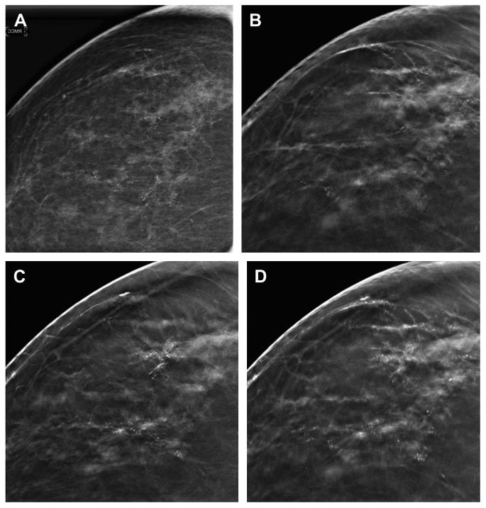
Fig. 11.

Slabbing to aid in the analysis of calcifications. A two-dimensional CC DM spot magnification view (A) shows suspicious calcifications in the lateral breast. A 1-mm reconstructed DBT slice in the CC projection (B) shows some of the lateral, linear calcifications but unsharpness of other calcifications. A different, 1-mm reconstructed DBT slice in the CC projection (C) shows additional calcifications that are now in-plane and in focus in an area of subtle architectural distortion. The calcifications seen on the previous slice are not as clearly visible on this 1-mm reconstructed slice. A 10-mm reconstructed “slab” (D) better demonstrates the extent of the suspicious calcifications and associated subtle distortion. The slabbing technique may help improve the conspicuity of a larger area of calcifications but also introduces a degree of unsharpness as the reconstruction thickness is increased. On biopsy, this was high-grade ductal carcinoma in situ without invasion.
