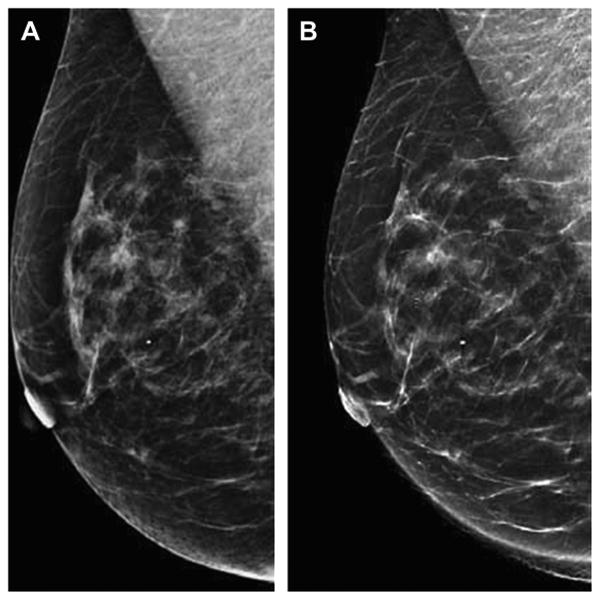
Fig. 12.

Synthetic two-dimensional images reconstructed from DBT acquisition. The two-dimensional DM MLO view is shown on the left (A) with the reconstructed synthetic MLO view (B) shown on the right (B). The synthetic image (B) is reconstructed by summing the data obtained from the individual slices that make up the DBT image set. Research and development is ongoing to reconstruct two-dimensional images that provide the necessary two-dimensional information, such as the morphology and distribution of clinically significant calcifications, so that two-dimensional DM imaging and the associated dose could be eliminated in many cases. The synthetic two-dimensional image would be viewed with the DBT image set.(Courtesy of Hologic, Inc, Bedford, MA; with permission.)
