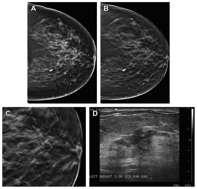
Fig. 2.

Malignancy detected on DBT only. (A) This patient has scattered fibroglandular densities and no abnormality was detected on the DM imaging. (B) The CC DBT view shows an area of architectural distortion in the retroareolar plane. (C) An enlarged, cropped view of the DBT in-plane slice of the area of distortion demonstrates the greater conspicuity of the area on tomosynthesis imaging. (D) An ultrasound image clearly shows an irregular mass with ductal extension. On biopsy, this was an invasive ductal carcinoma.
