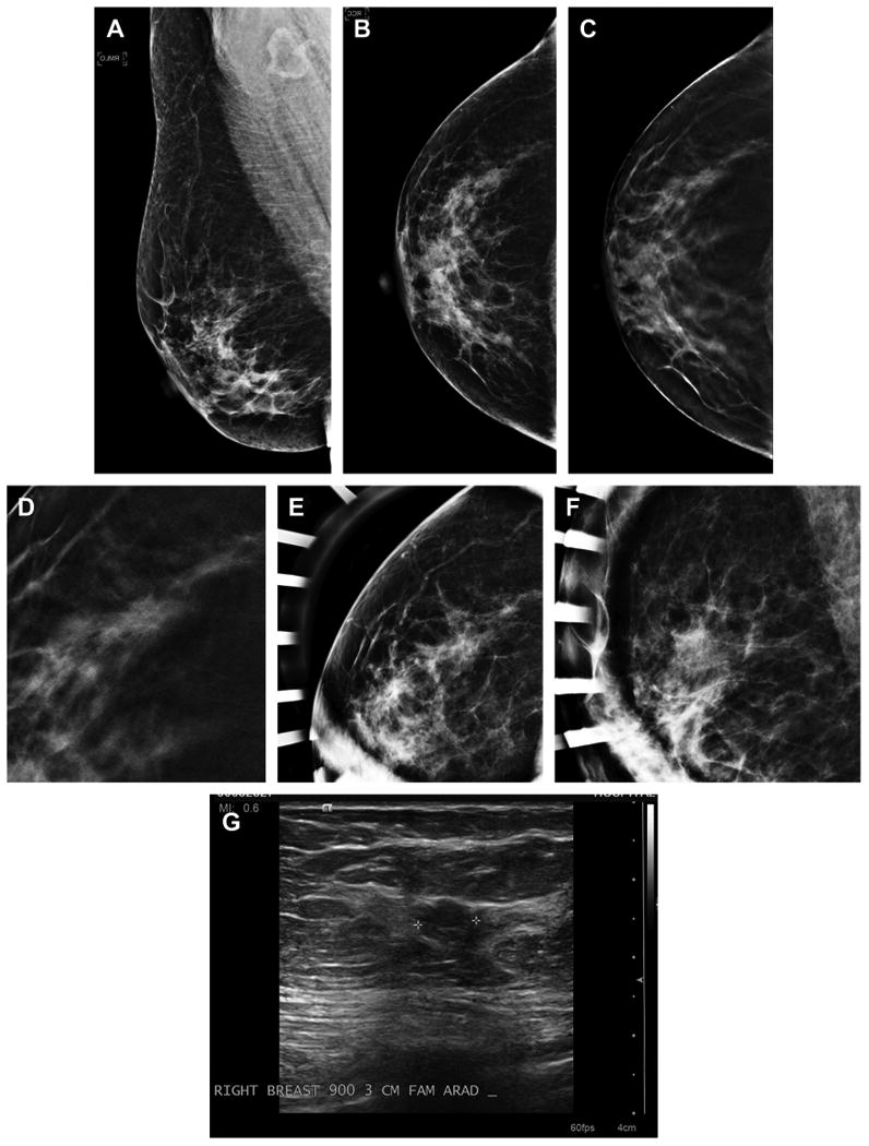
Fig. 3.

Cancer seen on only one view of DBT. A 54-year-old woman with normal MLO (A) and CC (B) two-dimensional mammography has very subtle spiculated mass seen in the lateral breast on the DBT CC view only (C). An enlarged, cropped view of the in-plane DBT slice where the subtle speculated mass was detected is shown (D). The patient was brought back from screening and additional spot magnification views were performed in the CC (E) and MLO views (F). There was no definite mass or distortion seen on the diagnostic two-dimensional imaging but on ultrasound (G) an irregular mass was visible in the area detected on the CC DBT view. An invasive ductal carcinoma was found on biopsy.
