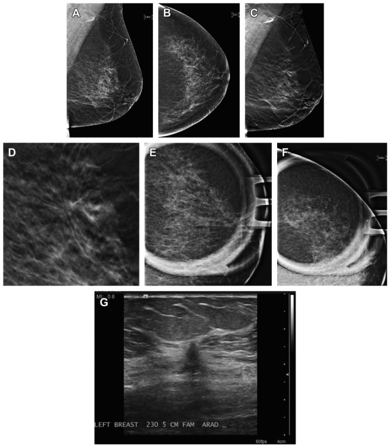
Fig. 4.

Cancer seen on MLO DBT view only. A 66-year-old woman presented for screening and has normal two-dimensional DM MLO (A) and CC (B) views. Note that the breast has very little glandular tissue to obscure lesions. On the DBT MLO view (C) a subtle area of distortion is present in the superior breast. An enlarged, cropped view (D) of the MLO in-plane DBT slice clearly shows the distortion. Spot magnification two-dimensional views in the MLO (E) and CC (F) views fail to show a discrete mass or persistent area of distortion. Ultrasound (G) was performed based on the three-dimensional localization from the MLO and ML DBT image set. A small, 5-mm intermediate grade invasive ductal carcinoma was found on biopsy.
