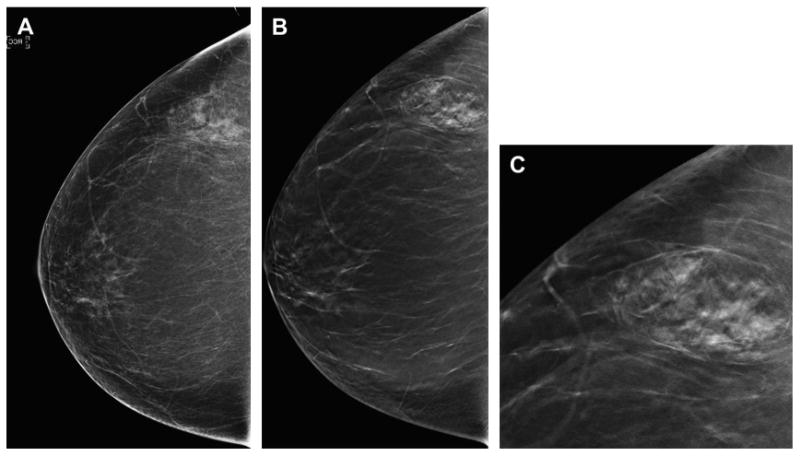
Fig. 6.

Benign lesion more conspicuous on DBT. A 53-year-old woman presents for baseline screening and has almost entirely fatty breasts except for focal asymmetry in the lateral right breast on the DM CC view (A). On the CC DBT series (B) the lesion is clearly a hamartoma; therefore, no further imaging is needed. An enlarged, cropped view (C) from the CC DBT series clearly shows the mixed density lesion with a pseudocapsule, typical of a hamartoma. No further imaging was needed and the patient was returned to routine screening.
