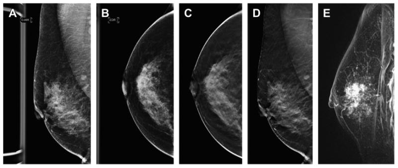
Fig. 7.

DBT shows the extent of malignancy better than two-dimensional DM imaging. This patient presented for screening and on the DM MLO view (A) no abnormality was seen. On the DM CC view (B) there was a subtle approximately 1.5-cm area of distortion seen in the lateral breast. Both the DBT CC and MLO series (C, D) show extensive distortion caused by a large mass in the superior subareolar location, much more conspicuous than the subtle, small area seen on the DM CC view. A contrast-enhanced breast MR image (E) shows a similar extent of disease as that seen on the DBT study. The patient had a 5-cm invasive ductal carcinoma with an extensive in situ component.
