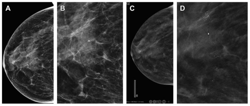
Fig. 9.

Three-dimensional localization of skin calcifications with DBT. The two-dimensional DM CC view (A) shows multiple clusters of calcifications. An enlarged, cropped two-dimensional CC image (B) shows calcifications that are not clearly benign. The CC DBT image (C) from the last, inferior or caudal, reconstructed slice (C) shows that all the calcifications are localized within the skin. Note the location graphic in the left corner of the image that shows that the slice is the first slice in the series (Slice: 1/46), at the “F” foot or caudal portion of the stack of DBT reconstructed images. Also visible are small round areas of lucency at the edges of the image. These are the caves of Kopans, columns of fat that extend from the dermis to the subcutaneous tissue. These are also seen on the magnified CC DBT view (D) again confirming that the calcifications are clearly within the skin and are therefore benign. No additional imaging is needed.
