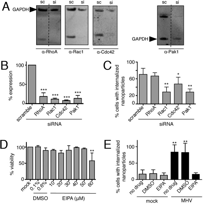
FIG 3 .

MHV-induced macropinocytosis is dependent on the classical macropinocytosis pathway. (A, B) Cells were reverse transfected with siRNA for 72 h, and protein knockdown was confirmed by immunoblotting (A) and standardized to GAPDH (B). Scrambled siRNA (sc)- and siRNA (si)-treated samples are from the same gel for each protein. RhoA and Pak1 are from discontinuous lanes separated by dashed lines. Data are represented as the means ± the standard errors of the means in triplicate assays. (C) Cells were reverse transfected for 68 h and infected with MHV for 8 h. Nanoparticles were added during the final 3 h, and cells were washed, fixed, stained, and imaged. Data are represented as the means ± the standard errors of the means of two replicates performed in duplicate, n = ≥30 fields per replicate. (D) The 12-h toxicity of EIPA was assessed with CellTiter-Glo. (E) Cells were mock infected or infected with MHV at an MOI of 1 PFU/cell for 8 h with no drug, DMSO, or 40 µM EIPA. Nanoparticles were added during the final 3 h of infection. Cells were washed, fixed, stained, and imaged, and the percentage of cells with internalized nanoparticles was calculated. Data are represented as the means ± the standard errors of the means of two replicates performed in duplicate. Significance was assessed by one-way ANOVA with Dunnett’s post hoc test. *, P < 0.05; **, P < 0.01; ***, P < 0.0001.
