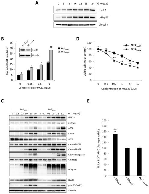
Figure 1. Hsp27 overexpression inhibits apoptosis and unfolded protein response induced by MG132 in PC-3 cells.
A, PC-3 cells were treated with 5μM MG132 for the indicated time. Protein levels were analyzed by western blotting. B Hsp27 overexpressing stable PC-3 (PC-3Hsp27) cells and control vector-transfected (PC-3Empty) cells were treated with indicated concentration of MG132 for 24 hours and apoptotic rates (subG0/G1 fraction) were quantified using flow cytometry. C, PC-3Hsp27 and PC-3Empty cells were incubated with indicated concentration of MG132. ER stress, ubiquitination and apoptosis were analyzed by changes in UPR and UPS markers expression, and caspase-3 and PARP cleavage using western blotting. D, PC-3Hsp27 and PC-3Empty cells were treated with indicated concentration of MG132 for 24 hours and cell growth was determined by crystal violet assay. E, Proteasome activity was monitored in PC-3Hsp27 and PC-3Empty cells and PC-3 parental for cleavage of the fluorescence substrate Suc-LLVY-AMC. Fluorescence was quantified using a spectrofluorometer (Fluoroskan Ascent FL, Thermo Labsystem). Bars, SD. ** differ from control (P < 0.05).

