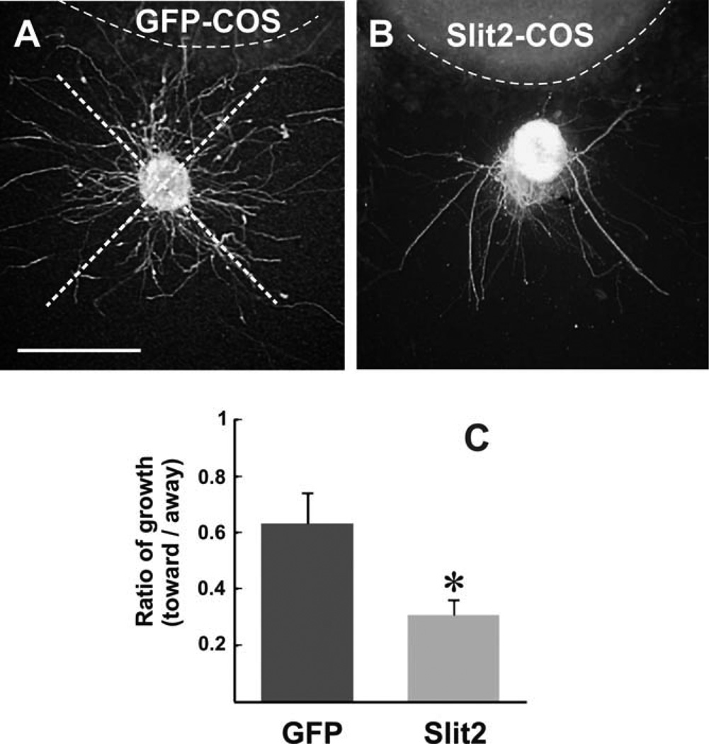
Fig. 3.
Slit2 inhibits TPOC axon growth in vitro. A,B: Explants from E10.5 embryos containing the nTPOC cultured in collagen gels exposed to COS cell aggregates transfected with a Slit2-myc (B) or a GFP expression vector (A) followed by β-tubulin III immunostaining. C: Quantification of neurite outgrowth within quadrants facing toward or away from the COS cell aggregate as shown in A. Graph shows the average ratio of toward/away growth (±SEM; n = 10 explants for GFP, n = 9 explants for Slit2). Explants had less neurite growth toward Slit2 aggregates than to control aggregates (*P < 0.05 by t-test). Scale bar = 100 µm.

