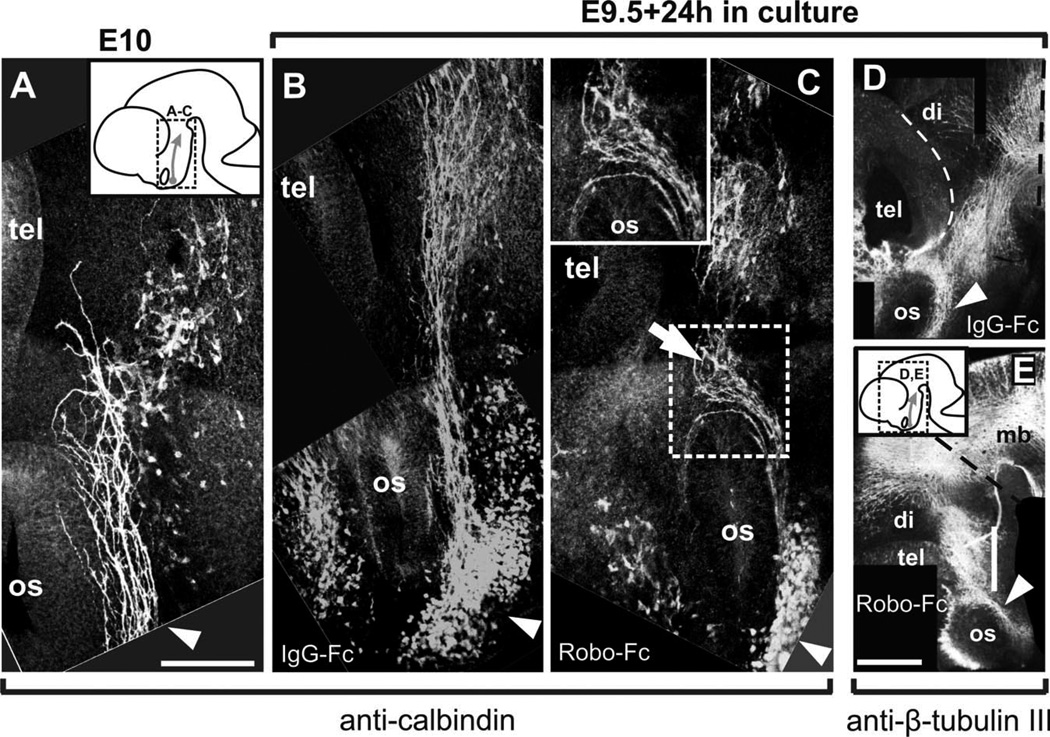
Fig. 4.
Impairing Robo function in cultured embryos causes alterations to TPOC projection. Calbindin immunostaining of the brain of E10 embryos allows visualization of the early stages of TPOC projection (A). E9.5 embryos were cultured for 24 hr in the presence of IgG-Fc protein (B) or in the presence of Robo1 and Robo2 Fc chimeras (C), followed by calbindin immunostaining. Approximate location of the brain region shown in A–C is indicated in the inset in A. The brains of embryos cultured with Fc control proteins (D) or with Robo-Fc chimeras (E) were immunostained for β-tubulin III. Region shown in D and E is indicated in the inset in E. tel, Telencephalon; mb, midbrain; hb, hindbrain; di, diencephalon; os, optic stalk. Scale bars = 100 µm in A (applies to A–C); 200 µm in E (applies to D,E).

