Fig. 5.
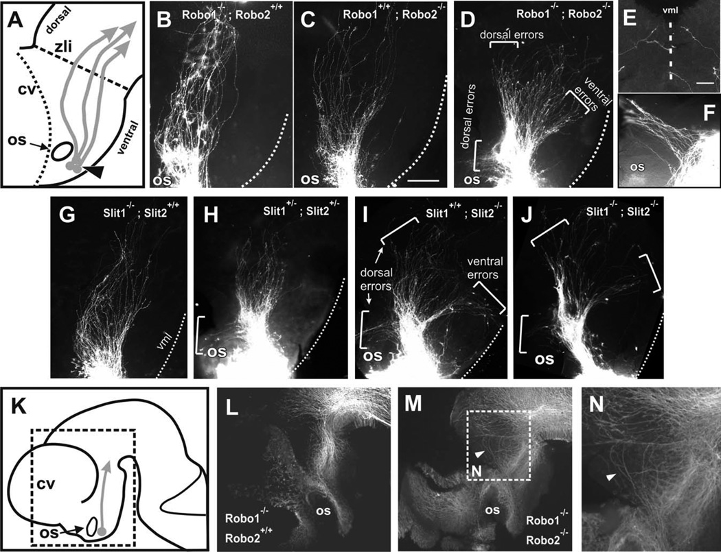
Loss of Robo1-Robo2 and of Slit1-Slit2 results in TPOC path-finding errors. DiI labeling of the TPOC in E10.5–E11 mouse embryos. A: Diagram of anterior forebrain (rostral is to the left) showing the normal TPOC trajectory (arrows) and the location of the DiI crystals for TPOC labelling (arrowhead). B,C: Robo1 and Robo2 single mutants have largely normal TPOC trajectories, although Robo2 single mutants display deviations limited to a slight dorsal widening of the tract (C). D: In Robo1/2 double mutants, TPOC axons make severe pathfinding errors, resulting in misprojection (brackets) or in expanded tracts. E: Ventral midline view of a Robo1/2 double mutant brain showing bilaterally labelled axons aberrantly invading the ventral midline. F: High magnification of an embryo showing axons turning dorsally around the optic stalk. G: Slit1−/− mutants appear largely normal. H: Slit1/Slit2 double heterozygotes have some axons turn (bracket) around the optic stalk, while most axons continue normal trajectories. I: Slit2 mutants have a wider TPOC and axons wander both ventrally and dorsally (brackets; these errors were seen bilaterally in two of three analyzed of this genotype). J: Slit1/Slit2 double mutants have extensive projection errors, similar to Slit2 and Robo1; Robo2 mutants. Control (L) and Robo1/Robo2 double mutants (M,N) were immunostained for β-tubulin III to reveal potential alterations to tracts other than the TPOC. Location of regions shown in L and M is indicated in K. Location of N is indicated in M. cv, Cerebral vesicles; os, optic stalk; zli, zona limitans intrathalamica; vml, ventral midline. Scale bars = 100 µm in C (applies to B–D,G–J); 200 µm for L,M; 50 µm in E (applies to E,F).

