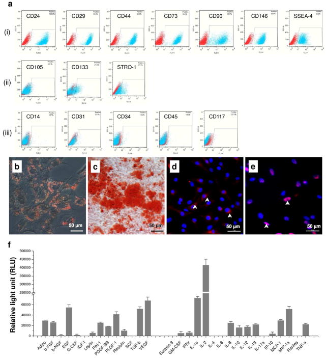
Fig. 1.
Characterization of hUSCs. (a) Cell surface marker expression of hUSCs was detected by flow cytometry. Strongly positive markers i: CD24, CD29, CD44, CD73, CD90, CD146, and SSEA-4; weakly positive markers ii: CD105, CD133, and STRO-1; and negatively expressed markers iii: CD14, CD31, CD34, CD45, and CD117. Multi-differentiation potential of hUSCs in vitro was evaluated using osteogenic, adipogenic, and myogenic induction. Osteogenic differentiation: (b) Alizarin Red S staining for calcium deposition ; adipogenic differentiation: (c) Oil Red O staining for lipid droplets ; and myogenic differentiation: immunofluorescent staining (arrows, red) for myogenic marker expression (d): Desmin and (e): MyoD in induced cells. Scale bar = 50 μm. (f) Trophic factors secreted by hUSCs were analyzed by human cytokine ELISA plate array and were mainly categorized as growth factors and immunomodulatory cytokines.

