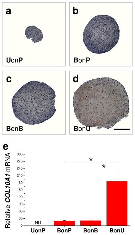
Fig. 4.
Hypertrophic evaluation of expanded cells. After expansion on Plastic, BECM, or UECM, hBMSCs or hUSCs were cultured in a pellet system supplemented with a serum-free chondrogenic medium for 14 days. (a–d) Immunohistochemical staining (IHC) was used to detect type X collagen. Scale bar = 800 μm. (e) TaqMan® real-time PCR was used to evaluate COL10A1 mRNA. * p < 0.05. Data are shown as average ± SD for n = 4.

