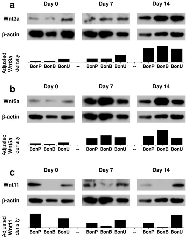
Fig. 5.
Involvement of noncanonical Wnt signaling pathway during chondrogenic induction. The pellets from hBMSCs expanded on three substrates were used to extract proteins for gel running and western blot. The pellets from hUSCs after expansion on Plastic were too tiny to provide sufficient protein for this measurement. Canonical [(a) Wnt3a] and noncanonical Wnt [(b) Wnt5a and (c) Wnt11] signaling pathways were evaluated in chondrogenically differentiated hBMSCs at day 0 (condensation phase), day 7 (early phase of chondrogenic induction), and day 14 (late phase of chondrogenic induction). β-actin served as an internal control. Image J software was used to semi-quantify immunoblotting bands.

