
Figure 6.
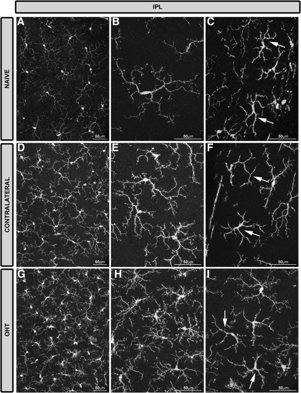
Iba-1+ cells in the IPL after 15 days of unilateral laser-induced OHT. Iba-1 immunostaining. Retinal whole-mounts. Most Iba-1+ cells in the IPL were ramified and there were few Iba-1+ cells with a dendritic-like appearance (arrow in C, F, I). In comparison with naïve eyes (A-C), ramified Iba-1+ cells in the IPL of contralateral eyes and OHT eyes formed a denser cellular mosaic (D, E, G, H). Both ramified and dendritic-like Iba-1+ cells exhibited morphological signs of cell activation (process retraction and increased secondary and superior order processes) that were more pronounced in OHT eyes (G-I) than in contralateral eyes (D-F). (IPL: inner plexiform layer; OHT: ocular hypertension).