
Figure 3.
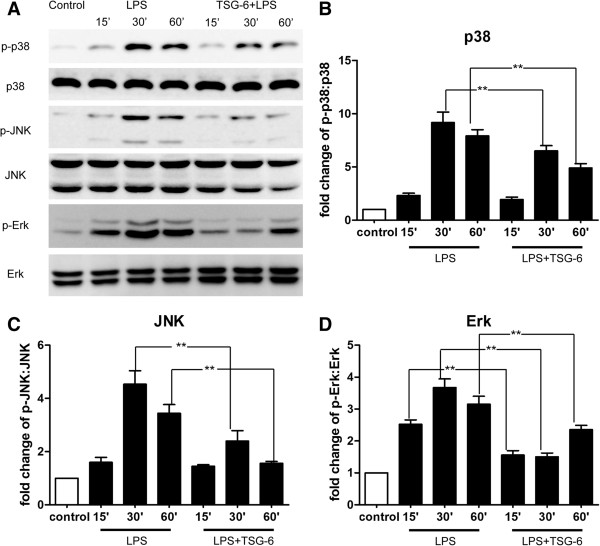
TSG-6 suppresses LPS-mediated phosphorylation of p38, JNK, and Erk. BV2 cells were stimulated with LPS in the presence and absence of rmTSG-6 (10 ng/ml) for the indicated times. Cell lysates were analyzed for the phosphorylated levels of p38, JNK, Erk, and their total levels by western blotting with the respective specific antibodies. (A) Representative image of protein levels of P38, JNK, and Erk. (B-D) The phospho:total ratios of p38 (B), JNK (C), and Erk (D) were determined by measurements of band intensities of each protein kinase and quantified as fold changes over the control (unstimulated BV2 cells). X axis are shown in time slots of 15, 30, and 60 minutes. Values are mean ± SD. n = 3; **P <0.01; significantly different from LPS-treated cells. Abbreviations: LPS, lipopolysaccharide.