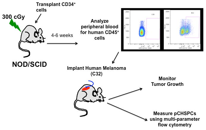
Figure 2. Schematic of C32 Melanoma Xenograft Model.

Mice were sub-lethally irradiated with 300 rads and human CD34+ cells were then transplanted into NOD/SCID mice. Following 4 weeks of engraftment, peripheral blood was analyzed for human CD45 and then C32 melanoma cells were implanted on the flanks of the humanized mice with tumor growth and CHSPCs both being monitored.
